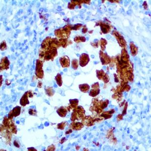
IHC of Cytokeratin HMW/AE3 on a FFPE Salivary Gland Tissue
| Intended Use | For In Vitro Diagnostic Use | |||||||||||||||||||||||||||||||||||
| Summary and Explanation | Cytokeratins are intermediate-filament keratins found in the intracytoplasmic cytoskeleton of epithelial tissue. There are two types of cytokeratins: the low-weight, acidic Type I cytokeratins and the high-weight, basic or neutral Type II cytokeratins. Cytokeratins are usually found in pairs comprising a Type I cytokeratin and a Type II cytokeratin. Expression of these cytokeratins is frequently organ or tissue-specific. Cytokeratin, High Molecular Weight AE3 (HMW, CK 8 ) is capable of recognizing all basic cytokeratins; therefore, it is a broadly reactive antibody staining most epithelia and their neoplasms. Cytokeratin HMW/AE3 stains normal and neoplastic cells of epithelial origin. CK HMW is primarily found in the non-squamous epithelia and is present in the majority of Adenocarcinomas and Ductal Carcinomas. It is absent in Squamous Cell Carcinomas. Hepatocellular Carcinomas are defined by the use of antibodies that recognize only cytokeratin 8 and 18. | |||||||||||||||||||||||||||||||||||
| Antibody Type | Mouse Monoclonal | Clone | AE3 | |||||||||||||||||||||||||||||||||
| Isotype | IgG1 | Reactivity | Paraffin, Frozen | |||||||||||||||||||||||||||||||||
| Localization | Cytoplasmic | Control | Prostate, Bladder, Salivary Gland | |||||||||||||||||||||||||||||||||
| Presentation | Cytokeratin HMW AE3 is a mouse monoclonal antibody derived from cell culture supernatant that is concentrated, dialyzed, filter sterilized and diluted in buffer pH 7.5, containing BSA and sodium azide as a preservative. | |||||||||||||||||||||||||||||||||||
| Availability |
| |||||||||||||||||||||||||||||||||||
| Note: For concentrated antibodies, please centrifuge prior to use to ensure recovery of all product. | ||||||||||||||||||||||||||||||||||||