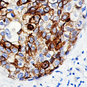
IHC of Myobacterium Tuberculosis on an FFPE Infected Lung Tissue
| Intended Use | For Analyte Specific Reagent | |||||||||||||||||||||||||||||||||||
| Summary and Explanation | Mycobacterium tuberculosis is a pathogenic bacterial species of the Mycobacteriaceae family and the causative agent of most cases of tuberculosis. M. tuberculosis has an unusual, waxy coating on its cell surface (primarily due to the presence of mycolic acid), which makes the cells impervious to Gram staining; M. tuberculosis can appear Gram negative and Gram positive in clinical settings. The Ziehl-Neelsen stain, or acid-fast stain, is used instead. M. tuberculosis is highly aerobic and requires high levels of oxygen. Humans are the only known reservoirs of M. tuberculosis. When in the lungs, M. tuberculosis is taken up by alveolar macrophages, but they are unable to digest and eradicate the bacterium. Its cell wall prevents the fusion of the phagosome with lysosome, which contains a host of antimycobacterial factors. Antibiotic resistant strains of mycobacterium tuberculosis have developed resistance to more than one TB drug, due to mutations in their genes. M. tuberculosis is characterized by caseating granulomas containing Langhans giant cells, which have a “horseshoe” pattern of nuclei. Cells are often seen wrapped together, due to the presence of fatty acids in the cell wall that stick together. This appearance is referred to as chording, like strands of chord that make up a rope. The clinical and histological criteria used to diagnose lymphadenitis caused by Mycobacterium tuberculosis complex organisms have poor specificity. Acid-fast staining and culture have low sensitivity and specificity. The diagnosis of tuberculosis by immunohistochemistry can be used to detect the mycobacterial antigen on formalin-fixed tissue biopsies and it’s consider fast, sensitive, and a highly specific method for establishing the etiological diagnosis of tuberculosis in histologic specimens. | |||||||||||||||||||||||||||||||||||
| Antibody Type | Rabbit Polyclonal | Clone | Polyclonal | |||||||||||||||||||||||||||||||||
| Isotype | IgG | Reactivity | Paraffin, Frozen | |||||||||||||||||||||||||||||||||
| Localization | Cell Wall | Control | Infected Tissue | |||||||||||||||||||||||||||||||||
| Presentation | Mycobacterium tuberculosis is a purified immunoglobulin fraction of rabbit antiserum that is filter sterilized and diluted in buffer pH 7.5, containing BSA and sodium azide as a preservative. | |||||||||||||||||||||||||||||||||||
| Availability |
| |||||||||||||||||||||||||||||||||||
| Note: For concentrated antibodies, please centrifuge prior to use to ensure recovery of all product. | ||||||||||||||||||||||||||||||||||||