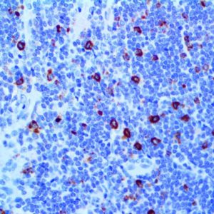
IHC of CD79a on an FFPE Colon Tissue
| Intended Use | For In Vitro Diagnostic Use | |||||||||||||||||||||||||||||||||||
| Summary and Explanation | CD79a is non-covalently associated with membrane-bound immunoglobulins on B-cells to constitute the B-cell Ag receptor. CD79a first appears at pre B-cell stage and persists until the plasma-cell stage, where it is found as an intracellular component. CD79a is found in the majority of Acute Leukemias of precursor B-cell type, in B-cell lines, B-cell Lymphomas, and in some Myelomas. CD79a is a B-cell marker that is generally used to complement CD20. This antibody will stain many of the same Lymphomas as CD20, but also stains more B-precursor Lymphoid Leukemias than CD20. CD79a also stains more cases of Plasma-cell Myeloma and occasionally some types of endothelial cells as well. CD79a will stain many cases of Acute Promyelocytic Leukemia (FAB-M3), but only rarely stains other types of Myeloid Leukemia. | |||||||||||||||||||||||||||||||||||
| Antibody Type | Mouse Monoclonal | Clone | JCB117 | |||||||||||||||||||||||||||||||||
| Isotype | IgG1/K | Reactivity | Paraffin, Frozen | |||||||||||||||||||||||||||||||||
| Localization | Membranous | Control | Tonsil, Lymph Node | |||||||||||||||||||||||||||||||||
| Presentation | CD79a is a mouse monoclonal antibody derived from cell culture supernatant that is concentrated, dialyzed, filter sterilized and diluted in buffer pH 7.5, containing BSA and sodium azide as a preservative. | |||||||||||||||||||||||||||||||||||
| Availability |
| |||||||||||||||||||||||||||||||||||
| Note: For concentrated antibodies, please centrifuge prior to use to ensure recovery of all product. | ||||||||||||||||||||||||||||||||||||